TOPIC DETAILS

  Team Member Role(s) Profile
Paul Banaszkiewicz Paul Banaszkiewicz Section Editor, Segment Author
Zak Zakareya Gamie Segment Author
  • The cell cycle is divided into four active phase and one inactive (quiescent) phase.
    • G0
  • Resting state
    • G1
  • Initial growth phase. Cells increase in size, produce RNA and synthesise protein.
    • S phase
  • DNA synthesis
  • Replication of DNA occurs during the S phase, so that the nucleus in G2 (4N) (tetraploid) contains twice the amount of DNA present in G1 (2N) (diploid).
    • G2
  • As the formation of DNA is an energy draining process, the cell undergoes a second growth and acquisition stage.
  • The cell continues to grow and produce more proteins.
  • The duplicated chromosomes are segregated and distributed into two daughter nuclei.
  • The M phase has several distinct phases:
  • Prophase

The chromatid coils and supercoils, making it more compact, the nuclear envelop dissolves.

  • Metaphase

This begins when the chromosomes have reached their maximal contracture. They move to the equatorial plate of the cell and the spindle forms.

  • Anaphase

This is triggered when a large protease called separase becomes activated following destruction of its inhibitory regulator. Each centromere divides and the new chromosomes begin to move towards opposite poles of the spindle.

  • Telophase

The chromosomes reach the poles of their respective spindles, the nuclear envelope reforms, chromosomes uncoil into chromatin form, and the nucleolus reforms.

  • Cytokinesis

This is the process where one cell splits off from its sister cell. Whereas mitosis is the division of the nucleus, cytokinesis is the splitting of the cytoplasm and allocation of the golgi, plastids and cytoplasm into each new cell.

  • Interphase is the stage in the life cycle of a cell where the cell grows and DNA is replicated.
  • Each chromosome has its own pattern of DNA synthesis, and some segments replicate early (e.g. hours).
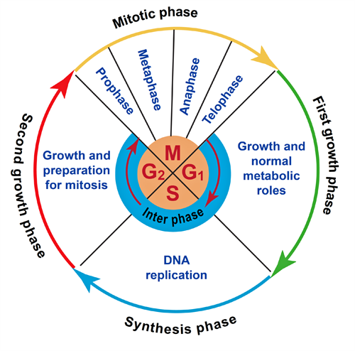
BS8CELLBIOLOGY1Harry.png

Figure 1. The cell cycle

  • In the cell cycle, there are three major points at which the cell “checks” itself before moving on.
  • Adequate energy reserves, size and damage to DNA are evaluated at the G1 checkpoint. If conditions are inadequate, the cell will halt the cycle and not continue to the S phase of interphase. Growth factors play an important role in carrying the cell past the G1 checkpoint.
  • The G2 checkpoint ensures all the chromosomes have been replicated and that the replicated DNA is not damaged. If the DNA has been correctly replicated, cyclin dependent kinases(CDKs) signal the beginning of mitotic cell division. Radiation therapy and chemotherapy can cause blockage of the G2 check point.
  • The M check point occurs near the end of the metaphase stage of mitosis. It determines whether all the sister chromatids are correctly attached to the spindle microtubules before the cell enters the irreversible anaphase stage.

BS8CELLBIOLOGY2.png

Figure 2. Internal checkpoints in the regulation of the cell cycle

  • Various proteins regulate the cell cycle.
  • A Cdks is an enzyme that adds negatively charged phosphate groups to other molecules in a process called phosphorylation. Through phosphorylation, Cdks signal the cell that it is ready to pass into the next stage of the cell cycle.

Cyclins

  • Cyclins undergo a constant cycle of synthesis and degradation during cell division. When cyclins are synthesised, they act as an activating protein and bind to Cdks forming a cyclin–Cdk complex. This complex then acts as a signal to the cell to pass to the next cell cycle phase. Eventually, the cyclin degrades, deactivating the Cdk, thus signaling exit from a particular phase. There are two classes of cyclins: mitotic cyclins and G1 cyclins.
  • The retinoblastoma protein (pRb)is a tumour suppressor protein that prevents excessive cell growth.
  • It blocks the growth of dysfunctional cells between the G1 and S phase of the cell cycle before they can undergo DNA replication.
  • pRb also regulates the function of cyclin and cyclin-dependent kinase (CDK) complexes which are needed for the cell to proceed with cell growth.
  • E2F is a transcription factor that forms a complex with pRb to inhibit cell growth. When E2F is not bound to pRb it encourages cells to proliferate, its main function is to express genes required for DNA replication to take place.
  • Retinoblastoma protein is dependent on the transcription factor E2F, which is bound to pRb's active site when it is hypophosphorylated. In tumorous cells pRb is dysfunctional .If there is a mutation in this gene, its binding domain is dysfunctional and can lead to excessive cell growth, which may cause tumour development.
  • Mutated pRb has been found in many human tumours, such as osteosarcoma and retinoblastoma.
  • The phases where pRB and E2F function are necessary and are during the Go phase, and during the restriction point. In these phases pRb will inhibit cell growth, and determine if the cell can transition from the G1 phase to the S phase.
  • If the cell is not growing adequately, for example if its DNA is damaged, then the retinoblastoma protein will hypophosphorylate and bind to the E2F transcription factor. This triggers the inhibition of E2F and of cyclin CDK complexes, and pRb is activated to stop cell growth. When pRB is phosphorylated, it no longer inhibits cell growth and the cell can continue developing. Phosphorylation of retinoblastoma is caused by the cyclin CDK complexes that are active throughout the cell cycle.

BS8CELLBIOLOGY3.png

Figure 3.pRb/EF binding in the cell cycle


  • Tumour protein p53 is a gene that codes for a protein that regulates the cell cycle and hence functions as a tumour suppression. It is very important for cells in multicellular organisms to suppress cancer. P53 has been described as “the guardian of the genome,” referring to its role in conserving stability by preventing genome mutation.
  • It is located on chromosome 17. If P53 is damaged tumour suppression is greatly reduced. As many as 50% of all human tumours contain p53 mutants.
    • It can activate DNA repair proteins when DNA has sustained damage.
    • It can arrest growth by holding the cell cycle at the G1/S regulation point on DNA damage recognition (if it holds the cell here for long enough, the DNA repair proteins will have time to fix the damage and the cell will be allowed to continue the cell cycle).
    • It can initiate apoptosis (i.e. programmed cell death) if DNA damage proves to be irreparable.

Apoptosis

  • Programmed cell death. Death from the inside out.
  • Requires a series of intracellular signalling events.
  • Stimuli that promote or inhibit apoptosis have the capacity to deregulate normal growth and differentiation.
  • Organised cascade of intracellular processes leading to the digestion of cellular DNA and fragmentation of the cell into apoptotic bodies.

Necrosis

  • Cell death from the outside.
  • Stem cells represent unspecialised cells that have the ability to differentiate into diverse specialised cell types and self-renew to produce more stem cells.
  • The two main types of stem cells are:
  1. Embryonic stem cells
  2. Adult stem cells
  • Other types of stem cell include:
  1. Foetal stem cells
  2. Amniotic stem cells
  3. Induced pluripotent stem cells (IPSCs)
  4. Nuclear transplant stem cells (ovasomes)
  5. Parthenote stem cells
  • By definition a stem cell requires that it possess two properties:
  1. Self-renewal: the ability to go through numerous cycles of cell division while remaining undifferentiated. If stem cells could not self-renew, tissues would run out of replacement cells for those that had died.
  2. Potency: the capacity to differentiate into specialised cell types. This requires stem cells to be either totipotent or pluripotent – to be able to give rise to any mature cell type.
  • There are two types of division:
  1. Symmetrical division: This allows the stem cell to self-renew and create more stem cells.
  2. Asymmetrical division: This produces a stem cell and a progenitor cell that will eventually differentiate into a particular cell.
  • Totipotent cells have the capacity to form an entire organism as well as the extraembryonic tissue including the placenta. Embryonic cells within the first couple of cell divisions after fertilisation are the only cells that are totipotent.
  • Pluripotent cells have the capacity to differentiate into any cell type in the body. They give rise to most, but not all of the tissues necessary for fetal development.
  • Multipotent cells can develop into more than one cell type, but are more limited in capacity than pluripotent cells. They can form many types of cell in a given lineage, but not cells of other lineages.

Progenitor cells

  • A progenitor cell is a biological cell that, like a stem cell, has a tendency to differentiate into a specific type of cell, but is already more specific than a stem cell and is pushed to differentiate into its “target” cell.
  • Progenitors are cells at a stage in between stem cells and mature functioning cells. The most important difference between stem cells and progenitor cells is that stem cells can replicate indefinitely, whereas progenitor cells can divide only a limited number of times.

Embryonic stem cells

  • Embryonic stem cells are pluripotent having the capacity to differentiate into any cell in the body.
  • They are only found in the early developmental stages of an organism (inner cell mass of a blastocyst) and represent the only cell type that has the ability to renew itself indefinitely.
  • As a unique precursor cell, it can differentiate into cells of all three germ layers: mesoderm, endoderm or ectoderm.

Adult stem cells

  • Adult stem cells are multipotent (lineage restricted) with a more limited capacity to differentiate. They are found throughout the body after development and are responsible for maintaining and repairing the tissue they reside in.
  • They show limited differentiation potential to tissues of one germ layer. Although a variety of multipotent adult stem cells exist, mesenchymal stem cells (MSCs) are probably the most interesting to orthopaedic surgeons because of their potential to differentiate into both bone and cartilage.

Fetal stem cells

  • Fetal stem cells can be isolated from fetal blood and bone marrow, other fetal tissues such as liver or kidney, or extra embryonic structures of fetal origin.
  • They are less ethically contentious than embryonic stem cells and their differentiation potential appears greater than adult stem cells.
  • Foetal blood is a rich source of both haemopoietic stem cells (HSCs) and non-haemopoietic MSCs. Fetal HSCs and MSCs have advantages over their adult counterparts, including greater multipotentiality and lower immunogenicity.
  • Fetal stem cells do not form teratomas.
  • Advantageous features include relatively easy accessibility and high proliferation rate.

Amniotic stem cells

  • Amniotic fluid is a source of multipotent stem cells.

Induced pluriotent stem cells

  • Induced pluripotent stem cells (iPSCs) are a type of pluripotent stem cell that can be generated directly from adult somatic cells that normally cannot differentiate at all. In 2006 Yamanaka et al. demonstrated that the introduction of four specific genes encoding transcription factors (proteins that switch on genes) could genetically convert adult cells into pluripotent stem cells.
  • Because iPSCs are made from a patient’s own cells – they avoid any issues with immune rejection. In addition there is a lack of ethical implications as cells are harvested from consenting adults rather than embryonic cells.
  • These patient-specific cells are the basis of an emerging technology can be used to study diseases in vitro, to test drugs on a human model without causing harm, and possibly to act as tissue replacement for diseased and damaged cells.
  • Although iPS cells make excellent laboratory models for studying diseases they have as yet to be implemented in human trials.
  • At present iPS cells have a high oncogenicity due to the transcription factors used for dedifferentiation, which are known to increase greatly the risk of tumours developing. Initially retroviruses were used that carried target DNA to be inserted into a host cell’s genome on injection, making them ideal for incorporating the four genes into the target cells. However this DNA and the rest of the virus’s genomes remain in the host genome, which can lead to transcription of unwanted genes and greatly increases the risk of tumours developing. This was a significant barrier to their use. A number of non-retroviral methods of iPS cell production have subsequently been developed.
  • Excisions

The excision strategy (transient transfection) of iPSC generation allows the transgenes to integrate briefly into the genome but then removes them once reprogramming is achieved.

  • Adenoviral methods

Unlike retroviruses, adenoviruses do not incorporate their genome into the host DNA. Because the transgenes are never incorporated into the host’s genome; they do not have to be excised. Instead the genes are expressed directly from the virus genome and are a safer method successfully to re-engineer cells.

Nuclear transfer stem cells (ntES cells)

  • Nuclear transplantation refers to the process of replacing egg chromosomes with the chromosomes of another cell.
  • These stem cells arise from eggs that have not been fertilised by sperm. Instead the DNA-containing nucleus has been removed from the cell and replaced with the DNA from a somatic adult cell (usually skin cell). This DNA is now encorporated into the egg. The egg is able to remodel the transferred genes and start dividing into multiple cells so that it can support early embryo development from the cleavage to blastocyst stage. This technology offers the opportunity to create patient-specific rejection-proof cells and tissues for transplantation. ntES cells are thought to provide the most genetically pristine source for creating genetically.

Parthenote stem cells (pES cells)

  • Parthenote stem cell are derived from eggs artificially activated without sperm to a dividing state.
  • An egg ready to be fertilised is activated by chemicals or a small electrical jolt. The activated egg divides into smaller and smaller cells. At approximately 100 cells, the cells on the outside form a sealed layer that surrounds a fluid-filled cavity with a group of cells inside (blastocyst). The inside cells can be removed to a petri dish and will continue to divide into parthenote stem cells.
  • Although slightly less versatile than ntES cells they are technically less difficult to produce.

Mesenchymal stem cells

  • Although all types of stem cells may have potential uses in orthopaedics, ethical and legal concerns have limited the use of embryonic stem cells while safety issues have restricted the use of induced pluripotent stem cells.
  • Orthopaedic surgeons have therefore focused their attention on mesenchymal stem cells (MSCs).
  • MSCs are multipotent stromal cells of mesodermal origin that show good differentiation potential towards several connective tissue cell types that include osteocytes, chondrocytes, adipocytes, tenocytes and myocytes.
  • MSCs are found in and can be harvested from a number of mesenchymal tissues including bone marrow, fat, synovial membrane, peripheral blood and periosteum.
  • MSCs can be isolated from bone marrow aspirates by their ability to adhere to plastic culture plates, a technique that allows them to be separated from most of the other cellular components of the marrow that do not adhere.1 However, these cells consist of a heterogeneous population of cells including mixed MSC clones at various stages of differentiation together with contaminant mononuclear cells and fibroblasts. They require additional processing.
  • Bone marrow-derived stem cells are harvested via a needle through a bone marrow aspirate. Although adipose tissue is a rich source of MSCs, fat-derived MSCs have consistently underperformed bone marrow-derived cells.

Definition of MSCs

  • Due to the lack of specific mesenchymal cell markers and the heterogeneity of the MSC populations, the International Society for Cellular Therapy (ISCT) has established three minimal criteria to define MSCs isolated from human bone marrow and other mesenchymal tissues:
  1. Plastic adherence
  2. Specific surface antigen (Ag) expression
  3. Multilineage potential
  • First, MSCs must be plastic-adherent when maintained in standard culture conditions using tissue culture flasks. Second, ≥95% of the MSC population must express CD105, CD73 and CD90, as measured by flow cytometry. In addition, these cells must lack expression (≤2% positive) of CD45, CD34, CD14 or CD11b, CD79a or CD19 and HLA class II. Third, the cells must be able to differentiate to osteoblasts, adipocytes and chondroblasts under standard in vitro differentiating conditions.
  • In recent years, stem cells have become a focus of regenerative medicine. Adult stem cells, harvested directly from bone marrow, adipose tissue or blood have the ability to undergo mitosis as well as multipotent differentiation into a variety of cell lineages.
  • ·         The goal of stem cell therapy is to replace or replenish diseased tissue through the localised differentiation of transplanted stem cells into cells which advance the healing process or directly restore the tissue physically.

Bone fractures and non-unions

  • Stem cells may stimulate bone growth and promote healing of fractures. Traditionally, bone defects have been treated with solid bone graft material placed at the site of the fracture or non-union. Stem cells and progenitor cells are now placed along with the bone graft to stimulate and speed the healing.

Articular cartilage damage/degeneration

  • Current techniques to treat articular cartilage damage use grafting and transplantation of cartilage to fill the defects. It is hoped that stem cells will create growth of primary hyaline cartilage to restore the normal joint surface.

Ligaments and tendon injury/degeneration

  • Mesenchymal stem cells may also develop into cells that are specific for connective tissue. This would allow faster healing of ligament and tendon injuries, such as quadriceps or Achilles tendon ruptures.

Spinal cord injury2

  • There has been much recent research into cell-based therapies for spinal cord injury. May be able to limit cell death, stimulate axonal growth, and replace injured cells.
  • Adult stem cells, Schwann cells, and olfactory ensheathing cells lack the ability to differentiate into cells of the central nervous system.
  • Mesenchymal stem cells can decrease cell death by modifying the local environment into which they are introduced. They prevent the activation of T-cells and thus downregulate the immune response accountable for the secondary mechanism of injury after spinal cord trauma. They also have neurotropic properties secreting nerve growth factor and neurotrophin-3, which promotes axonal growth.
  • Multiple animal studies have shown that ESCs can differentiate into neural progenitor stem cells of the CNS, can limit the inflammatory response and cell death associated with spinal cord injury and have the ability to stimulate axonal growth. Although a small number of phase I and II trials have taken place and show promising results to date no significant high quality published results exist on the use of ESCs in humans. A significant drawback is that ESCs stimulate the immune response.
  • Animal studies with induced pluripotent stem cells suggest benefits similar to those offered by ESCs without the ethical considerations surrounding use of the latter cells, and that because the autologous transplantation of iPSCs is possible, there is no need for immunosuppression with their use.

Meniscal injury

  • Isolated case reports exist of meniscal regeneration after percutaneous injection of autologous ASCs into an adult human knee.3 It is not clear whether this is a direct action of the mesenchymal-based cells or is rather mediated by secretion of certain stimulating factors on the existing meniscal tissue.

Intervertebral disc disease

  • The causes of disc degeneration are both complex and multifactorial. An imbalance between extracellular matrix degradation and synthesis results in progressive collapse and mechanical failure of the disc. An overall decrease in resident disc cell number and function with cellular responses leads to alterations in both the cartilaginous and proteoglycan matrix components of the disc.
  • Numerous clinical trials have been undertaken using MSCs biologically to repair degenerative disc. Percutaneous stem cell-mediated disc degeneration has the potential to establish itself as a possible treatment option in patients with low back pain hoping to avoid invasive spinal surgery.

Spinal fusion

  • The vital elements in bony fusion are an adequate quantity of bone-forming cells (osteogenesis), an appropriate microenvironment directing bone synthesis through a variety of growth factors (osteoinduction), and a scaffold or cage in which the growth of bone is well positioned (osteoconduction).
  • Pseudoarthrosis remains a pressing issue occurring in 13–41.4% of patients undergoing spinal fusion. Risk factors include older age, thoracolumbar kyphosis, smoking, diabetes mellitus, metabolic bone disease and female gender.
  • MSCs and ADSCs have both demonstrated a significant positive effect on spinal fusion in a number of experimental models. Cellular in vitro expansion is necessary to increase the number of viable pluripotent cells along with the addition of growth factors and/or BMPs.

Physeal injury/defects

  • Several animal models have investigated the use of stem cells combined with growth factors to promote the regeneration of damaged regions of the growth plate.

Duchenne muscular dystrophy

  • Duchenne muscular dystrophy (DMD) is a progressive and incurable severe muscle-degenerative disease caused by a mutation in the dystrophin gene. The dystrophin protein helps strengthen and connect muscle fibres and cells.
  • Induced pluripotent stem cells (iPS cells) have the ability to become any type of human cell while also maintaining the genetic code from the person they originated from. Scientists have removed the Duchenne mutations in the iPS cells using a gene editing platform. The iPS cells free from the Duchene mutation can then be transplanted back as skeletal muscle cells into recipients.

Osteogenesis imperfecta

  • OI is a rare genetic disorder caused by a mutation in the COL1A1 and COL1A2 genes in chromosomes 17 and 7. This mutation results in the production of the osteoblasts of a qualitatively and quantitatively defective type I collagen.
  • Stem cell therapy would aim to replace abnormal osteoblasts, the bone forming cells with defective bone matrix production with normal functioning osteoblasts. Some small clinical trials and isolated case studies have shown a transient improvement in growth velocity.

Juvenile arthritis

  • MSCs play a role in the modulation of host immune response by inhibiting the proliferation of T lymphocytes.
  • The use of MSCs is appealing in diseases in which there is an increased T-cell response such as inflammatory arthritis.

Avascular necrosis of the femoral head

  • Several clinical trials with implantation of stem cells into the necrotic area report improved femoral head survival compared to controls.
  • MSCs have been reported to promote tumour growth and metastases. There is very limited clinical experience with pluripotent stem cells (embryonal stem cells and iPSC). Based on their features of self-renewal and high proliferation rate the risks of tumour formation should be considered high.
  • Ethical issues and controversies regarding the use of embryonic stem cells and embryos exist.
  • Donor site morbidity from stem cell harvesting.
  • Processing time (requires separate procedures to harvest and re-implant cells).
  • Retroviruses may be used to generate human iPSCs. These viruses are genetically altered to encode the genes that are required for transformation into an iPSC. Applying this genetic reprogramming, the used viruses can integrate into the cell genome. Consequently the cells may contain multiple viral integration sites in their genomes.
  • Cost
  • Microbial contamination during cell amplification.
  • Controlling stem cell differentiation into the desired cell lineage.
  • Control of their proliferation and differentiation into complex, viable 3D tissues is challenging.
  • Lack of adequate vehicles/scaffolds for implantation of stem cells.
  • Integration with local tissues.
  • Immunological rejection and disease transmission (if allogeneic).
  • Potential modulation of host immune system by implanted stem cells.
  • Continuous cell amplification of MSCs may lead to chromosomal abnormalities.

Table 1. Overview of risk factors and risks associated with stem cell-based therapy4


Risk factors or hazards

Identified risks

Intrinsic factors

Origin of cells (e.g. autologous vs. allogenic, diseased vs. healthy donor/tissue)

Rejection of cells

Cell characteristics

Differentiation status

Disease susceptibility


Tumorigenic potential

Unwanted biological effect (e.g. in vivo differentiation in unwanted cell type)


Proliferation capacity

Toxicity


Life span

Neoplasm formation (benign or malignant)


Long term viability


Excretion patterns (e.g. growth factors, cytokines, chemokines)


Extrinsic factors manufacturing and handling

Lack of donor history

Disease transmission


Starting and raw materials

Reactivation of latent viruses


Plasma-derived materials

Cell line contamination (e.g. with unwanted cells, growth media components, chemicals)


Contamination by adventitious agents (viral/bacterial/mycoplasma/fungi, prions, parasites)

Mix-up of autologous patient material


Cell handling procedures (e.g. procurement)

Neoplasm formation (benign or malignant)


Culture duration


Tumorigenic potential (e.g. culture-induced transformation, incomplete removal of undifferentiated cells)


Non-cellular components


Pooling of allogenic cell populations


Conservation (e.g. cryopreservatives)


Storage conditions (e.g. failure of traceability, human material labelling)


Transport conditions


Clinical characteristics

Therapeutic use (i.e. homologous or non-homologous)

Undesired immune response (e.g. GVHD)


Indication

Unintended physiological and anatomical consequences (e.g. arrhythmia)


Administration route

Engraftment at unwanted location


Initiation of immune responses

Toxicity


Use of immune supressives

Lack of efficacy


Exposure duration

Neoplasm formation (benign or malignant)


Underlying disease


Irreversibility of the treatment


  • Blastocyst: The 4–9-day-old embryo (post-fertilisation), which consists of 100–200 total cells. This stage of development is prior to implantation in the uterus. Only two types of cells are present at this time, the trophectoderm (foundation of the placenta) and the inner-cell mass, which will also contribute cells to the extraembryonic tissues as well as the entire fetus.
  • Inner cell mass (ICM): A population of cells (approximately 20–30%) at the blastocyst stage that generate certain extraembryonic cells and tissues as well as those of the entire embryo.
  • Epigenetic: Chemical modifications of DNA that do not alter a gene’s sequence, but impact gene expression.
  • Episome: A stable DNA molecule that persists in the nucleus without integrating into the cellular genome.
  • Genome: The entire set of genetic material in an organism.
  • Genotype: The DNA sequence of a gene.
  • Mesenchymal stem cells: A specific class of adult or tissue-specific stem cells that generates connective tissues (including cartilage, tendons, and bone).
  • Mesoderm: One of the three basic embryonic germ layers which includes blood, muscle, and bone.
  • Transduction: The introduction of genetic material into a cell using a viral vector.
  • Transcription: The process of “reading” a gene to make an RNA “message” that is then translated into a protein.
  • Transfection: The process of introducing new genetic material into a cell. Various methods are used: some rely on physical treatment (electroporation, cell squeezing, nanoparticles, magnetofection), others on chemical materials (calcium phosphate, liposomes) or biological particles (viruses) that are used as carriers.
  • Transcription factor: A protein that binds to specific DNA sequences, thereby controlling the rate of transcription of genetic information from DNA to messenger RNA. Transcription factors perform this function alone or with other proteins in a complex, by promoting (as an activator) or blocking (as a repressor) the recruitment of RNA polymerase.
Previous
Next

References

  • 1. Oehme D, et al. Cell-based therapies used to treat lumbar degenerative disc disease: a systematic review of animal studies and human clinical trials. Stem Cells International 2015; 2015: 16.
  • 2. Schroeder GD, Kepler CK, Vaccaro AR. The use of cell transplantation in spinal cord injuries. J Am Acad Orthopaed Surg 2016;
  • 3. Pak J, Lee JH, Lee SH, Regenerative repair of damaged meniscus with autologous adipose tissue-derived stem cells. BioMed Res Int 2014; 2014.
  • 4. Herberts CA, Kwa M, Hermsen H. Risk factors in the development of stem cell therapy. J Transl Med 2011; 9(1): 29.