TOPIC DETAILS

  Team Member Role(s) Profile
Fazal Ali Fazal Ali Section Editor
Yuraj Yuvraj Agrawal Segment Author

Document title: Examination of the Hip
Section editor: Mr Fazal Ali
Segment author: Mr Yuvraj Agrawal
Document history: Examination of the hip

For an exit fellowship examination of the hip not only must candidates be able to examine the hip well but they must also ensure the eaminers see and apprciate each part of their examination technique.

It will be immediately apparent is a candidate has developed a routine with their examiantion approach.

Pain

  • Groin, thigh and buttock pain typically indicates a hip pathology
  • Is exacerbated by activity and relieved by rest
  • Associated limited walking distance
  • Night sleep disturbances is a god indicator of severity of symptoms

Stiffness

  • Patients do not complain of stiffness but have difficulty with activities of daily living as a consequence of the stiffness.
  • Typically, difficulty with going up or down stairs, clipping their toe nails, putting socks and shoes and getting in and out of car etc.

Limping

  • It may or may not be associated with pain
  • Can be due to variety of reasons
  • Common reasons are limb length discrepancy, muscle weakness and bone and joint deformity

Past medical history

  • History of previous problem with their hips and surgery to the hip must be explored
  • History of long term steroids
  • History of treatment for cancer, HIV

Social History

  • Occupation
  • Type of accommodation
  • Use of walking aids
  • Hobbies and expectations
  • History of alcohol abuse and smokin

Expose the patient adequately and preferably wearing a pairs of shorts. Remove coats and cardigans. Check if they are comfortable and whether they require to use their walking aids for standing or walking.

  1. Stand the patient 
  2. Walk the patient
  3. Trendelenburg test  
  4. Lie the patient down & square the pelvis
  5. Thomas’s test and Range of movements  
  6. Limb length assessment 
  7. Examination of the spine, knee and distal neuromuscular examination

Look 

  • From the front for any asymmetry in the coronal plane. 
  • Comment on the attitude of the limb, obvious discrepancy in the length or bulk of quadriceps (Video 1). 
  • From the side, for sagittal balance, kyphosis, exaggerated lumbar lordosis, scar on the lateral aspect of the hip and ask for any other scar you can't see, occasionally in the groin.
  • From the back for gluteal, hamstring and calf asymmetry. This is a good opportunity to have look at the back for scoliosis, evidence of previous lumbar spinal surgery and occasionally stigmata of neurofibromatosis.
Pitfalls: If the patient stands stooping forward or with knee flexed, ask them if they could stand upright and straighter. If they are able to stand straighter but flex their knee or straighten their knee but stoop forward, they have a subtle fixed flexion deformity which you could confirm later - “a dipping duck sign”.

 Gait

Ask patients if they need to use a walking aid to walk. 

Give clear instructions to the patients to walk to the table, window or the wall opposite and back to you.  

Observe and comment on the excessive swaying of the shoulders, truncal motion, dip of the hemi-pelvis, foot progression angle and the three rockers of the foot. You can keep it simple by commenting on whether there is a heel strike and toe off or not and talk about the cadence (Video 2,3).

Remember to look at patient's face to assess if this is painful. Comment on what you see. 


Video 1.Ask the patient to walk and observe gait

Trendelenburg test

Explain to the patient what are you asking him to do first giving clear instructions.  

There are different techniques of doing this. The reader is recommended to pick any one and practice it and do it proficiently. You are testing the side on which the patient is standing on.  

Author's preferred technique is with thumb resting on the anterior superior iliac spine (ASIS) and the hands on the hips. Ask the patient to rest his hands on your arms for reassurance as well as to detect a subtle weakness in the abductors (Figures1,2). 

Clinical hip 1.png

Figure 1.Trendelenburg test

Clinical hip 2.png

Figure 2.Trendelenburg test.Thumbs over ASIS fingers feeling for contraction gluteal muscles.

Look and feel for the rise or dip of the hemi-pelvis. You may do this standing, sitting on a chair (if available) or kneeling down. Some patients try and compensate by leaning over to the side being tested to bring the centre of gravity over the leg.

A test is negative (normal) if the hemi-pelvis on the non-tested side rises

A test is positive (abnormal) if the hemipelvis on the non-tested side dips below the horizontal level. 

This is a test of abductor function and hence consider the different components if positive. The components are -

(1) force (abductor muscles)

(2) lever arm (abductor muscle length and femoral neck length) 

(3) fulcrum (the hip joint)

The above palpation technique also provides you an opportunity to have a feel for the level of the ASIS. If the ASIS are at different heights, suggesting a limb length discrepancy, you may perform the block test for functional limb length assessment at this stage.

Squaring the pelvis

Patient's pelvis must be squared as the first manoeuvre. Confirm this by checking that the pelvis lies perpendicular to the couch. Pelvis must be also squared if directed to measure the limb length on its own.

Thomas’s Test

This test is performed for assessing any fixed flexed deformity of the hip joint.  

After warning the patient, slide your hand under the lower back and ask the patient to flex their opposite knee as high as they can. This should obliterate any lordosis they may have (Image 3). 

Assess the degree of flexion at the hip, by assessing the angle formed at the knee on the examined side. This indicates the degree of fixed flexion deformity at the hip joint. Check if patient could improve this by pushing their knee down to the couch. This is the true fixed flexion deformity. 

Clinical hip 3.png

Figure 3.Thomas's test:note the lack of full extension on obliterating the lumbar lordosis

Video 2.Make sure that the lumbar lordosis is completely eliminated.

Pitfalls: Examiner must check if the patient has a prosthetic joint on the opposite side prior to the test. Consideration must be made if the patient is known to have ankylosing spondylitis; hip or knee arthrodesis (Video 5). 

For a patient with ankylosing spondylitis, proceed as normal to as far as possible to obliterate any compensation. The lumbar lordosis need not be completely flattened.

For patients with a hip arthrodesis, lift the contralateral leg until the lumbar lordosis is flattened (Video 5). 

Range of movement

Assess the range of flexion actively and check if this could be improved passively. 

This could be followed by passively assessing the internal and external rotation in flexion as well as in extension. 

Check for abduction and adduction in extension. The opposite leg will have to abducted to assess the degree of adduction. 

Stabilise the pelvis with a hand on the opposite pelvis for abduction and the same side for adduction.

Flexion: 0-130 degrees; abduction: 0-45 degrees; adduction: 0-30 degrees; extension : 0-10 degrees.

Leg lengths 

Pre-requisites: Pelvis must be squared to the couch and the limbs placed in identical positions to avoid misreading. If one is unable to place the limb in identical position, the limb length may have to measured in segments (femur & tibia) using bony landmarks. 

True limb length is measured from a fixed bony prominence above the hip joint (by convention anterior superior iliac spine) to the ipsilateral medial malleoli using a measuring tape. If there is a limb length discrepancy, Galeazzi's test is performed to identify the short segment.

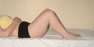
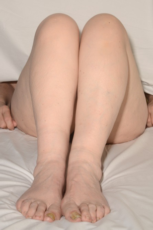
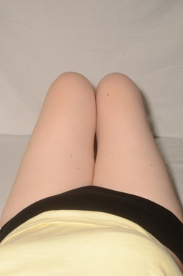
Galeazzi's test: Hips are flexed at 45 degrees, knees at 90 degrees and back of the heels are placed together (Image 4,5,6). 

Bryant’s triangle: If the asymmetry is identified in femoral length, Bryant's triangle is drawn to identify if the shortening is above or below the trochanter.

Apparent limb length is measured from a fixed central point, xiphisternum or umbilicus to medial malleoli and the lengths compared. This is measured to identify any pelvic obliquity - which may be fixed or compensatory.

Functional limb length is measured with patient standing on pre measured blocks. This relies on the patient's perception of the limb length discrepancy and planned correction.

Pitfalls: The shortening is sub-malleolar if there is a limb length discrepancy, but none detected on true or apparent limb length measurement. This is occasionally seen in patients with previous os calcis fracture or subtalar degeneration. Block test is recommended to quantify the limb length discrepancy.

If unable to place the limbs in identical position, measure different segments; femur and tibia from fixed and identical bony landmarks. Such a situation is encountered in patients with fixed genu valgum or varum
Clinical hip 4.png

Clinical hip 5.png

Clinical hip 6.png

Figures 4-6.Galeazzi's test

Pitfalls: The shortening is sub-malleolar if there is a limb length discrepancy, but none detected on true or apparent limb length measurement. This is occasionally seen in patients with previous os calcis fracture or subtalar degeneration. Block test is recommended to quantify the limb length discrepancy.

If unable to place the limbs in identical position, measure different segments; femur and tibia from fixed and identical bony landmarks. Such a situation is encountered in patients with fixed genu valgum or varum
Complete the examination with examination of spine, knee, pedal pulses and neurological examination distally.

Examination of a patient with arthrodesed hip

It is important to be aware of patients that have unusual features such as a fused contralateral hip or knee and be able to adjust examination technique as needed to take account of this 

Video 3.Examination patient ankylosed hip

Medical Illustration, Sheffield Teaching Hospital NHS Trust, Sheffield

Previous
Next

References

  • 1. Harris N & Ali F. Examination Techniques in Orthopaedics. Second edition, ed. Cambridge University Press 2014; 103-117.