TOPIC DETAILS

  Team Member Role(s) Profile
SattAR Sattar Alshriyda Section Editor, Segment Author
  • The foot exhibits both longitudinal (medial and lateral) and transverse arches.
  • These arches are maintained by: the shape of the foot bones, the activity of muscles and a wide variety of ligaments.
  • A flat foot (pes planus) is a foot with a large plantar contact area and a small or absent longitudinal arch in contrast to pes cavus (high arched foot).
  • Though flexible flatfeet are present in nearly all infants and many children the exact incidence is unknown.
  • It is present in 20-25% of adults and runs in families.

4.5.1.png

Footprint demonstrating the differences between normal, flat and high arched foot.

  • Though the cause is unknown it is associated with generalized ligamentous laxity and runs in families.
  • The diagnosis is clinical.
  • The medial arch reappears on tiptoeing or on passive dorsiflexion of the great toe (Jacks test).
  • The Achilles tendon may be tight but subtalar joint motion should not be limited.
  • Subtalar motion should be full.

4.5.2.jpg

Image source

Legend: Beightons assessment for ligamentous laxity

4.5.3.png

Flexible flat feet. On tip toeing, the heel moves into varus and the medial arch reconstituted. Bottom left picture shows Jacks test.

  • Plain radiographs are not required to make a diagnosis however they may be useful in quantifying the deformity
  • Several radiological parameters have been used to assess foot deformities.

On the lateral view:

  • The lateral distal tibial angle (LDTA=89°, range 86-92°). Abnormal value indicates distal tibia deformity which may contribute to the flat feet deformity.
  • The calcaneal pitch (17°, range 11-23°). The smaller the calcaneal pitch, the more plantarflexed the hindfoot.
  • The lateral tibiocalcaneal angle (69°, range 44-86°). The larger the tibiocalcaneal angle, the more plantarflexed the hindfoot.
  • The lateral talocalcaneal angle (49°, range 36-61°). The greater the talocalcaneal angle, the more valgus and abducted the hindfoot.
  • Lateral Meary’s angle (lateral talo-1st metatarsal angel) which is normally 5° (range 1-9°). The greater the lateral talo–first metatarsal angle, the more planus is the midfoot.

On the AP view

  • The anterior distal tibial angle (ADTA= 80°, range 78-82°).
  • The AP talocalcaneal angle of  Kite ( 21°, range 14-28°)
  • The AP  talo-first MT angle (10°, range -3-28°)

4.5.4.png

Lateral foot plain radiograph showing useful measurements to assess foot deformity. These are useful in assessing pes cavus and other deformities. (See also hallux valgus).

4.5.5.png

Lateral foot plain radiograph showing useful measurements to assess foot deformity. These are useful in assessing pes cavus and other deformities.

  • Treatment for a flexible flatfoot deformity is not required and the parents should be reassured.
  • Orthotic and operative treatment is controversial.
  • Orthoses (medial arch supports) have been shown not to promote the development of the longitudinal arch and should not be routinely prescribed (1).
  • They may however relieve pain if present.
  • Calf stretching exercises may be helpful if the Achilles tendon/gastrocnemius is tight.

Surgery

  • Surgery is reserved for the older child with intractable symptoms unresponsive to nonsurgical options.
  • A joint sparing operation-osteotomy such as a lateral column lengthening procedure with or without release of the gastrocnemius/Achilles tendon is the procedure of choice.
  • Triple C osteotomies ( Medial calcaneal slide, Cuboid opening wedge and medial Cuneiform dorsal opening wedge.
  • Arthroeresis of the subtalar joint (using an implant) is an option but is not without complication and the parents should be consented appropriately.
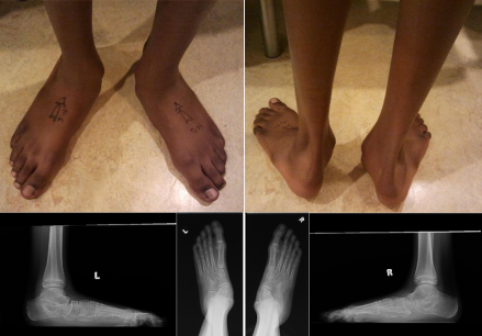
4.5.6.png

Clinical photograph and plain x-ray of a teenager with severe flat feet undergoing triple C osteotomy.
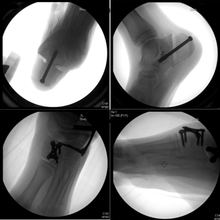
4.5.7.png

Fluoroscopy pictures of triple C osteotomies. Notice the medial calcaneal slide, Cuboid opening wedge and medical cuneiform dorsal opening wedge.
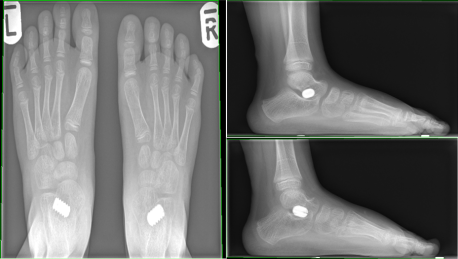
4.5.8.png

Arthrodesis for bilateral flat feet.

Previous
Next

References

  • 1. Gould N, Moreland M, Alvarez R, Trevino S, Fenwick J. Development of the child's arch. Foot Ankle. 1989 Apr;9(5):241-5.