TOPIC DETAILS

  Team Member Role(s) Profile
SattAR Sattar Alshriyda Section Editor, Segment Author
  • Tarsal coalition is an abnormal connection between 2 or more bones in the foot.
  • The connection may be fibrous, cartilaginous or bony.
  • The cause is unknown but failure of embryonic mesenchymal segmentation or differentiation or abnormal ossification has been postulated.
  • The reported incidence varies between 1 and 6 %.
  • There is a higher incidence among first degree relatives ( 39% has been reported).
  • Bilateral in 50%.
  • The commonest is calcaneo-navicular followed by talocalcaneal coalitions. Others are talo-navicular and calcaneo-cuboid.
  • They may be solitary or multiple ( hence CT or MRI scan is useful before surgery).
  • Coalitions are associated with limb abnormalities such as fibular hemimelia and proximal femoral focal deficiency.

4.6.1.png

Clinical photograph showing left rigid flat foot. Standing on toes cause the heel to move into varus and the medial arch is reformed, but this is not the case with the right foot.

  • Presents with pain and gait disturbance as coalition ossifies (Calcaneo-navicular age 8 - 12 and Talo-calcaneal age 12 – 16).
  • Peroneal and extensor tendons can be seen standing out on the lateral foot (hence the old name-peroneal spastic flat foot).
  •  Ankle movement is normal, but subtalar movements are restricted or absent.
  • Several signs on plain radiographs are very suggestive of tarsal coalition such as:
  1. Flatfeet
  2. Abducted forefoot
  3. Ant eater sign
  4. Ball and socket ankle joint
  5. C-sign
  6. Talar peaking
  • CT scan and MRI scan can confirm the diagnosis and rule other co-existing coalitions. They can also assess the adequacy of excision.
  • MRI scan can also showed local inflammation and bruises.
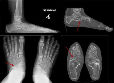
4.6.2.png

Plain radiograph and Ct Scan of a child with bilateral calcaneo-navicular tarsal coalition. Notice the flat foot on the lateral view and the forefeet abduction. Also noted the tarsal coalition marked with red arrow.
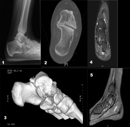
4.6.3.png

Plain radiograph and CT Scan of a child with left talo-calcaneum tarsal coalition. Notice the talar peaking on the lateral view. The CT Scan nicely shows the exact anatomy of the coalition. MRI scan showed high signal in the area indicating bruises or inflammation.

The goal of treatment is pain relief and to improve joint motion

Nonsurgical

  • Analgesia
  •  NSAIDs
  • Activity modification
  • Orthoses/insoles
  • Period of immobilization in a below knee weight bearing cast for 4-6 weeks.

Surgery

  • Calcaneonavicular coalition can be excised utilizing a modified Ollier incision.
  • Several materials are recommended to interpose to reduce the risk of recurrence.
  • Fat, tendon, bone wax are all used. None has been proven to be superior
  • Talocalcaneal coalitions may be excised if less than 50% of the middle facet is involved.
  • A medial hindfoot approach is used.
  • Excising the bar is unlikely to affect the associated deformity ( flat feet)
  • Residual deformity correction ( lateral column lengthening or triple C osteotomy may be undertaken as a second stage surgery or can be done at the time of excision (see flatfeet for further details)
  • Valgus heel of > 20° is a poor sign for bar excision   
  • A triple or subtalar arthrodesis may be required in the presence of degenerative changes, multiple coalitions or if symptoms persist despite previous surgery to excise a coalition.

4.6.4.png

Pre-operative (top) and postoperative (bottom) CT Scans showing adequate excision.

Previous
Next

References

  • 1. Gould N, Moreland M, Alvarez R, Trevino S, Fenwick J. Development of the child's arch. Foot Ankle. 1989 Apr;9(5):241-5.